In angle closure or narrow angle glaucoma, the iris physically obstructs the internal drain (trabecular meshwork) of the eye. This causes fluid build-up and elevated intraocular pressure which can lead to glaucoma. When a patient has narrow angles, fluid cannot move through the pupil normally, due to excessive contact between the iris and lens. Fluid builds up behind the iris, pushing it forward, further narrowing the angle and can lead to an acute angle closure attack (eye emergency) as well as the development of chronic angle closure glaucoma . This situation is known as pupillary block and is the most common cause of acute angle closure. LPI is a procedure that can be used to treat narrow angles.
When is LPI necessary?
Laser peripheral iridotomy is generally recommended for patients with narrow angles, narrow angle glaucoma, or acute angle closure glaucoma. In acute angle closure, LPI can be used to break the angle closure attack, lower the intraocular pressure, as well as prevent another attack of angle closure glaucoma. It is also helpful to widen the angle in patients with or at risk for chronic angle closure glaucoma, thus lowering their long term risk of glaucomatous damage and vision loss. If your physician determines that your angles are significantly narrow and you are at risk, LPI may be suggested as a treatment option.
Understanding the Laser Peripheral Iridotomy (LPI) Glaucoma Procedure:
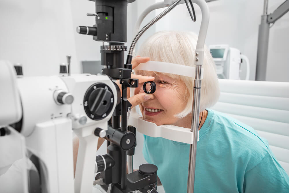
First, the eye is numbed with topical drops. An additional drop that constricts the pupil and thins out the iris as well as a glaucoma eye drop may be given before the laser as well. With a small lens, laser energy is then used to make a small microscopic hole in the iris (the colored part of the eye that controls the amount of light that enters into the eye).
This small opening allows fluid to now flow more freely from behind the iris into the anterior chamber and into the drain of the eye (trabecular meshwork). This helps alleviate pressure build up behind the iris, opens the angle, and allows the fluid to enter the drainage pathway more easily, resulting in a lowering of intraocular pressure.
