Glaucoma can be thought of as a characteristic change in the appearance of the optic nerve that is associated with a degradation of vision. Glaucoma is most commonly associated with an elevated eye pressure and loss of peripheral vision. The optic nerve is damaged in glaucoma. One can think of the optic nerve as a cable connecting the eyeball and the brain. The eyeball detects visual information. The optic nerve transmits the visual information, and the brain arranges this information into an image that we can recognize. If your optic nerve is damaged, your brain will receive incomplete information and may not be able to form an image that can be recognized or that is clear.
Glaucoma Defined
In simple terms, glaucoma is an eye condition where the eye pressure is elevated, causing damage to the optic nerve. Typically, “normal” eye pressure hovers around 21 mmHg and below. There are, however, special cases when the eye pressure is “normal” and yet the patient develops typical damage of the optic nerve that results in loss of peripheral perception and decreased vision. On the contrary, there are people who have persistently elevated eye pressure and suffer no damage to the optic nerve. Thus, we can see that glaucoma is not so simple. While elevated eye pressure is one of the primary risk factors, its presence or absence does not necessarily define whether a patient has glaucoma.
Glaucoma has been called the “silent thief of sight” because in most cases, the patient is not even aware that he or she has glaucoma. Fortunately, ophthalmologists can usually detect damage resulting from glaucoma before it negatively affects a patient’s vision. This is done by careful examination of the optic nerve along with specialized testing.
Causes of Glaucoma – Optic Nerve and Pressure
In the majority of cases, glaucomatous optic nerve damage occurs from elevated eye pressure. By lowering the eye pressure, most nerve damage can be stopped or slowed down so that the patient can live out their life with good sight. Glaucoma treatment is centered on the goal of lowering the eye pressure. This can be accomplished by using a combination of medicines, lasers and/or surgeries. Treatment plans are individualized for each patient to prevent or slow down further vision loss. In extremely rare cases, optic nerve damage continues even though the lowest eye pressure has been achieved. Intensive research in this area is now being performed to help understand the cause of the damage in these patients. New treatment paradigms are being investigated that may help preserve the optic nerve.
The pressure in the eye is directly related to the amount of fluid in the eye. The name of this fluid is aqueous humor. It is produced in the back portion of the eye and it drains in the front portion of the eye. When the amount of fluid made is equal to the amount of fluid drained, the eye pressure stays constant. Many conditions can cause an elevation in eye pressure such as aging, inflammation, bleeding, injury, tumors, or even birth defects can raise the eye pressure. Such conditions typically result in “clogged” or blocked drainage system. In most case of glaucoma, examination reveals a normal appearing eye and drainage system. In such cases, where no eye abnormalities are found, the condition is described as primary open angle glaucoma or “POAG”, the most common type of glaucoma. In other circumstances there is complete blockage or partial blockage of the drainage system. This is called angle-closure glaucoma. In other cases, there is “clogging” of the drainage system. There are many different types of mechanism that lead to the increase in the IOP, ultimately all damage the optic nerve.
“What is the status of my optic nerve?”
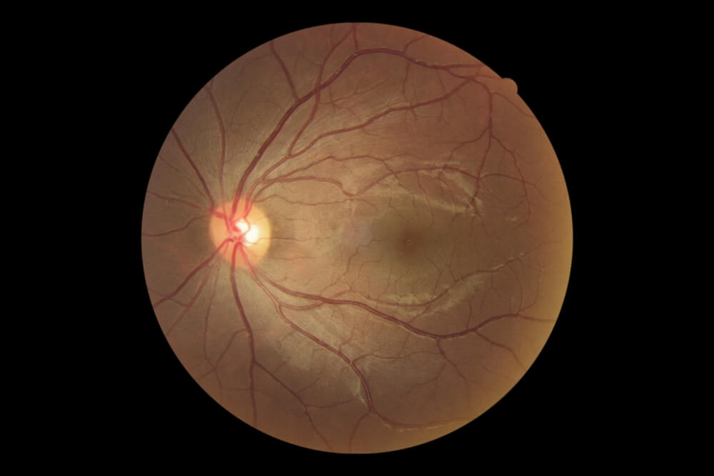
Normal optic nerve head 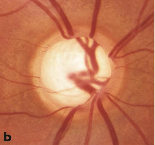
Glaucomatous Cupping
© 2022 American Academy of Ophthalmology
During your office visits, assessment of the optic nerve and its function will be carried out by direct examination, eye pressure measurements and specialized testing. Although many patients are rightfully concerned about the eye pressure measurements, it is the status of the nerve that is most important.
Optic Nerve Cupping in Glaucoma
The hallmark sign of glaucomatous damage is the change in appearance of the optic disc, or cupping. To understand optic disc cupping, we first have to understand how the body transmits information from the eye to the brain. Each eye is connected to the brain by an optic nerve, which on average is 3-4 mm wide. Inside this nerve there are around 1.2 million individual nerve cells, each of which is responsible for carrying visual information from a small and specific area of the visual field. If these nerve cells are damaged, for instance by glaucoma, then they can no longer carry information to the brain which results in blind spots. Damaged nerve fibers will tend to degenerate and “wither away”. There is an associated loss of support tissue, a reduction in blood vessels and a collapse in the support structure within the nerve. Together, the appearance of the optic nerve becomes more cupped and pale in uncontrolled glaucoma. See above for progression of cupping:
