Micro-invasive glaucoma surgery (MIGS)
Goniotomy (TrabEx® and Kahook Dual Blade Glide®)
Goniotomy is a well established surgical technique done using a single-use surgical device, such as the Kahook Dual Blade Glide® (KDB) and the TrabEx®. They both feature a dual-blade design, done as a stand-alone procedure or in conjunction with cataract surgery. When done with cataract surgery, it is commonly done after the cataract is removed by using the same clear corneal incision. Using a mirrored gonio lens for visualization, these devices are inserted into the anterior chamber of the eye and the trabecular meshwork is excised allowing better axis for the aqueous to schlemm’s canal and the collector channels. If successful, this results in better outflow and lower intraocular pressure. This procedure is minimally invasive and does not involve opening up the conjunctival tissue, making future glaucoma surgeries simpler if necessary.
Both TrabEx® and Kahook Dual Blade Glide® goniotomy are done in an ambulatory surgery center under local anesthesia and light intravenous sedation (like cataract surgery). In some cases an eye block may be done for additional anesthesia. Following this procedure you will be using post operative medications in addition to your glaucoma drops. If successful, you might be able to eliminate some of your glaucoma eye drops. Your surgeon will examine you the following day.
Hydrus® microstent
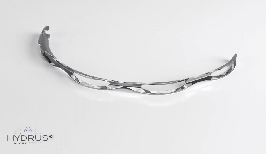
The Hydrus® microstent is an FDA-approved device implanted at the time of cataract surgery through the trabecular meshwork into schlemm’s canal to decrease intraocular pressure. The 8mm device is made of nitinol and works via bypassing the trabecular meshwork and dilating schlemm’s canal allowing better aqueous outflow and ultimately lower eye pressure. The device is implanted using the same incision used for the cataract extraction. This procedure is minimally invasive and does not involve opening up the conjunctival tissue, making future glaucoma surgeries simpler if necessary. Patients can safely undergo MRI in most circumstances.
If there are any questions regarding this please contact our office or visit the Hydrus® information page
The Hydrus® Microstent is done in an ambulatory surgery center under local anesthesia and light intravenous sedation (like cataract surgery). In some cases an eye block may be done for additional anesthesia. Following this procedure you will be using post operative medications in addition to your glaucoma drops. If successful, you might be able to eliminate some of your glaucoma eye drops. Your surgeon will examine you the following day.
Istent®
The iStent inject® W is an FDA-approved device implanted at the time of cataract surgery, using the same cataract incision. The newest device includes two titanium stents, less than 1mm, that are implanted through the trabecular meshwork into schlemm’s canal 2 to 3 clock hours apart to enhance aqueous outflow and lower intraocular pressure. This procedure is minimally invasive and does not involve opening up the conjunctival tissue, making future glaucoma surgeries simpler if necessary. Patients can safely undergo MRI in most circumstances. If there are any questions regarding this please contact our office or visit the Istent® information page.
The istent inject® W is done in an ambulatory surgery center under local anesthesia and light intravenous sedation (like cataract surgery). In some cases an eye block may be done for additional anesthesia. Following this procedure you will be using post operative medications in addition to your glaucoma drops. If successful, you might be able to eliminate some of your glaucoma eye drops. Your surgeon will examine you the following day.
GATT (Goniotomy Assisted Transluminal Trabeculotomy)
GATT is a minimally invasive glaucoma surgery performed using small micro-incisions in the cornea. After entering the eye, the trabecular meshwork is pierced, schlemm’s canal is then cannulated 360 degrees, and ultimately unroofed. This allows the whole drainage system of the eye to be targeted, allowing increased aqueous outflow and reduction of intraocular pressure. This procedure is minimally invasive and does not involve opening up the conjunctival tissue, making future glaucoma surgeries simpler if necessary.
GATT is done in an ambulatory surgery center under local anesthesia and light intravenous sedation (like cataract surgery). In some cases an eye block may be done for additional anesthesia. Following this procedure you will be using post operative medications in addition to your glaucoma drops. If successful, you might be able to eliminate some of your glaucoma eye drops. Your surgeon will examine you the following day.
Omni® (Canaloplasty/trabeculotomy)
The Omni® surgical system is a minimally invasive implant free procedure using a small microcatheter through a small corneal incision. It can be done as a stand alone procedure or in conjunction with cataract surgery. The device penetrates the trabecular meshwork, catheterizes Schlemm’s canal, dilates the canal and distal collector channels using viscoelastic (canaloplasty), and ultimately unroofs the trabecular meshwork (trabeculotomy). This results in lowering resistance of aqueous outflow at multiple sites of the drainage system, leading to lower intraocular pressure. This procedure is minimally invasive and does not involve opening up the conjunctival tissue, making future glaucoma surgeries simpler if necessary.
Omni® is done in an ambulatory surgery center under local anesthesia and light intravenous sedation (like cataract surgery). In some cases an eye block may be done for additional anesthesia. Following this procedure you will be using postoperative medications in addition to your glaucoma drops. If successful, you might be able to eliminate some of your glaucoma eye drops. Your surgeon will examine you the following day.
ECP (Endoscopic cyclophotocoagulation)
ECP is most often done in conjunction with cataract surgery using the same incision. Following the removal of the cataract, a small probe with a camera is inserted into the eye. The probe is connected to a laser with fiber-optic cables that allows the laser energy to be delivered to the ciliary body, which is the structure producing fluid and pressurizing the eye. As a result, it causes coagulation of the ciliary body and a reduction of intraocular fluid, leading to reduced eye pressure. The laser energy can be titrated to achieve light blanching of the ciliary processes, which is directly visualized on a monitor during the procedure. This portion of the surgery takes only a few minutes and should cause minimal if any pain. This procedure is minimally invasive and does not involve opening up the conjunctival tissue, making future glaucoma surgeries simpler if necessary.
ECP is done in an ambulatory surgery center under local anesthesia and light intravenous sedation (like routine cataract surgery). In some cases an eye block may be done for additional anesthesia. Following this procedure, you will be using postoperative medications in addition to your glaucoma drops. It may take 6-8 weeks before the final outcome of the procedure is known. If successful, you might be able to eliminate some of your glaucoma eye drops. Your surgeon will examine you the following day.
CPC (Transscleral cyclophotocoagulation)
Transscleral cyclophotocoagulation (CPC) is often done as a stand alone procedure to lower intraocular pressure. Like ECP it works by shutting down the cells in the eye that produce the intraocular fluid (ciliary body). It is generally used in cases of advanced glaucoma or eyes that have had previous glaucoma surgeries. It is done in an ambulatory surgery center generally under IV sedation and an eye block. Following anesthesia, the surgeon will place the probe on the surface of the eye near the limbus (cornea/sclera junction), and then apply the laser energy. Generally, the surgeon will treat approximately 20 to 30 laser spots, 360 degrees, avoiding sites of previous surgeries and sensory nerves. The laser energy is transmitted through the tissue and into the ciliary body, resulting in coagulation and decreased production of intraocular fluid to lower the eye pressure.
No incisions or cuts into the eye are necessary and the surgeon can easily titrate the laser energy and treatment intensity based upon the patient’s needs. This procedure can be repeated and the response may not be determined for a few weeks post-operatively. You will leave the surgery center with an eye patch and generally will start drops later that day or the following day. Your surgeon will examine you the following day to check your pressure.
Xen® Gel Stent
The Xen® Gel Stent is a microsurgical implant used to lower eye pressure in the setting of more moderate or advanced glaucomas. The stent bypasses the traditional outflow pathway, allowing fluid to get out of the eye, where it drains into the subconjunctival space (filtering bleb) and eventually is absorbed back into the body. It can be done as a stand-alone procedure or in conjunction with cataract surgery. The Xen® injector can implant the stent via a small corneal incision (internal approach) or externally through the sclera and into the anterior chamber. When done externally, the conjunctival tissue does need to be opened up similar to traditional glaucoma filtering surgeries. Similar to the traditional glaucoma filtering surgery trabeculectomy, the surgeon will use the anti-scarring medication mitomycin-C (MMC) or 5-Fluorouracil (5-FU) to try to prevent the body from scarring down the stent.
This increases the chances that the Xen® will remain patent and flowing, resulting in lower intraocular pressure.
The Xen® is done in an ambulatory surgery center under light intravenous sedation (like cataract surgery) and usually a local eye block. Following this procedure you will be using post-operative medications but initially will be off of your glaucoma drops. Your surgeon will examine you the following day. Like traditional filtering surgeries, your doctor will monitor you carefully for the first 4-6 weeks as your pressure can be variable in the early postoperative course. There is no contraindication for patients to undergo MRI with this device following their surgery.
